Tuesday, July 10, 2012

Sorry. Ian Katz should have been on today. Here is a case I saw recently in a young male with a history of a lesion on his abdominal wall which had only been there for 3 or at the most 5 years. He had some total body photography images taken when he was 15 years old. He is now 26.
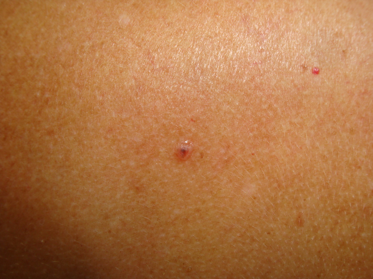
What do you think of this lesion? Happy to reassure him or would you cut it out? He has quite a few other clinically "atypical" nevi but none quite as large as this one.


Dr Cliff Rosendahl said...
Fantastic case with baseline and follow up images.
Chaos - absent. Leave!
A melanoma of this size would not be symmetrical!
This proves that 'congenital-type' naevi need not be present at birth.
Thanks Ian!

Dr Ian McColl said...
Glen, the advantage we have here is the total body photographs taken when he was 15 years old. There are none from the intervening time and he has not been regularly reviewed by anyone. This lesion was a standout in terms of it's size but is it symmetrical? Does it have any dermatoscopic clues to melanoma that would make you excise it?

Dr Alan Cameron said...
Great case Ian!

How do we weigh up the clear evidence of a new and enlarging lesion with a clue to melanoma (grey structures) and enough peripheral brown clods north and NW to at least raise the question of asymmetry against the strong impression of symmetry when viewed from a distance and the young age.

Banky et al found 126 changed and 155 new lesions in patients under 30 for a total of 4 melanomas. So around 70 benign for each melanoma.

Overall 18 melanomas were found using TBP in this study, only 4 were new lesions, 14 were changed. This is not surprising; there were more melanomas that were inconspicuous at initial photography that became apparent over a few years, than totally new ones.

Dr Ian McColl said...
I have put up the histology. Choose from Melanoma, Congenital nevus, Severely dysplastic compound nevus.

Dr Cliff Rosendahl said...
The histo is not a melanoma and not a congenital naevus, It's a compound naevus
Nests exceed single cells
Melanocytes mature as they descend (at least the very deep ones do)
It better be a benign compound naevus!

Dr Cliff Rosendahl said...
Keith the first histo slide has an interesting DE junction but can't assess well at low power. This is a great case and it may even provoke immuno-stains to assess maturity but on what I can see in those images I would think it is a compound naevus. I'll email Ian K and ask him to look

3:03 PM
Ian Katz said...
This is a real tough case histologically. That first slide does have quite a lot of flattening but there is also some scarring so that may be a regenerative area. The other histo slides show atypia in some areas but other areas resemble a congenital naevus.

Probably congenital naevus with scarring but would really have to examine carefully

Immunoperoxidase I find unhelpful in these cases

3:11 PM
Dr Ian McColl said...
OK Reported by my pathologist as severely dysplastic compound nevus. He felt the dermoepidermal junction changes were not those of a congenital nevus.
I see Cliff kept the dysplastic word out of his assessment but there is quite a bit of cytological atypia of those basal melanocytes and some but not all of the nests.
There was quite a bit of melaninin melanophages in the centre as well.

3:34 PM
Ian Katz said...
in other words it is a melanoma

Dr Ian McColl said...
I thought it was benign on dermatoscopic grounds when I saw him but he said this was still growing and changing by becoming darker centrally. The fact that it was such a standout and relatively new meant I really could not have left it. When patients say things are changing then unless i am 100% sure it is benign I usually excise. I did not need much urging to remove this one.

Harald Kittler said...
I changed my opinion after I had a closer look at the histopathology. First of all there is a banal nevus present for sure. No doubt about that! the fact that it was not present at age 15 doesn't mean anything. These type of nevi tend to appear in early childhood until late puberty and have a "congential pattern" on histopatology. The only question that remains is: IS there a melanom in situ in the center of the nevus or is this trauma related. In the absence of any convincing history of trauma you probably have to sign it out like melanoma in situ developing in a nevus. A limited lentiginous proferation of melanocytes at the junction is common over these types of nevi! This is probably too much (in the absence of trauma)
NEvi and MELANOMA are 2 different things and the fact that both may appear together does not mean they are part of a spectrum. I don not understand why this cannot be understood!
In sum: Most parts of this lesion are a banal nevus, no doubt,but a melanoma may develop in a banal nevus and this fact doesn't make the nevus "dysplastic" in retrospect.

5:35 PM
Harald Kittler said...
BTW: Melanoma in congenital nevi may develop anywhere (in the center like here or at the periphery), melanoma in Clark nevi usually develop at the periphery!

5:38 PM
Dr Cliff Rosendahl said...
Thanks harald and thank you for clarifying the insitu melanoma arising in naevus scenario. Do you think that there are dermatoscopic clues to the insitu melanoma? Do you think the appearance at the centre of this lesion relates to melanoma or is that all part of the dermatoscopic apprearance of the congenital naevus?

5:48 PM
Harald Kittler said...
Sure there are clues: Gray dots! the overall appearance is symmetric because the melanoma (if it is one!) happened to develop in the center of this lesion.

Harald Kittler said...
Gary, this case is different! This is not a nevus of the lentiginous type of the elderly. The content is always improtant in dermatscopy.

7:46 PM
Dr Cliff Rosendahl said...
This is a very good case Ian. We still don't know if there is melanoma there but it has made me aware that if melanoma arises in the centre of a congenital naevus there may be symmetry for a while. Clues need to be assessed in congenital naevi even in the absence of chaos!
Con should have had the honour but he is enjoying himself in Tahiti! I had intended putting up a case on an ear which I know is at least lentigo maligna but I have left the clinical image in a camera in my rooms! I did make a video of the histology which you can view below and I will put the clinical up first thing tomorrow morning. It was reported as a lentiginous dysplastic junctional nevus. Have a look at the video. Click the little box with the arrows pointing out to play it full screen and change the number for the resolution from 360 to 720. Press pause to let it load if your connection is slow. Press ESC on your computer to go back to your normal screen.


Also I wonder if you would tell the group of your favourite iPad Apps! Some of my favourites I have learned about from other people especially Jeff. Tell us your favourite or favourites and also why you think they are so good. They can be our Xmas present to each other.

Harald Kittler said...
Great video Ian. I completely agree with your interpretation!

1:24 AM
Marcia Sonneveld said...
Amazing presentation and, as an amateur have learnt heaps. Would love to see three histopath slides for comparison- the tru LDJN, this LM in situ and, an early invasive LM for comparison.Will watch again later.Trying to grasp why there is the lymphocytic reaction. Is it good or is it bad?

Blues revisited and a leg lesion

Some of the cases since we started back have been quite challenging! Three cases today that are back to basics. Case 2 might be a problem but everything is there and I have histology for all three.

Case 1, Lesion on the back. No previous history of skin cancers. Female aged 55.


case 2, Elderly male with this lesion on his lower back.


Case 3 Lesion on the lower leg in a male aged 65


Case 3


What would be your preferred diagnoses for these three lesions?

Dr Ian McColl said...
The histology for case 1 was no surprise. It was a BCC. I think the bit shown corresponds to that big blue clod. When I first looked at this clinically I thought it was a combined nevus but the dermatoscope helped.

Dr Ian McColl said...
Case 3 was a pig IEC. I have put up the histology of case 2. There are two ? lesions in the dermis and one at the dermepidermal junction. Cliff will work them out. Note the two different cell types in the dermal part. As far as I am aware nothing has previously been done to this lesion.

Dr Ian McColl said...
I have put up the histology of case 2 for Cliff.

Dr Cliff Rosendahl said...
I think I need a pathologist here! There is a proliferation of melanocytes at the DE junction with I guess a separate proliferation in the deep dermis of very mature looking melanocytes. ? Blue naevus with overlying invasive melanoma or reactivated naevus? The dermal part of the DE junction proliferation is not showing maturation. Not confident at all.

Dr Ian McColl said...
Well done Cliff. Case 2 was reported as a melanoma, clark level 2, 0.25 mm arising in association with a combined nevocellular nevus and a blue nevus. The blue nevus bit is the pigmented spindled cell area and the blue regular nevus cells in the dermis is the common nevus. The pathologist thought there was a bit of scarring above this deep cellular component and wondered if it was regression or if it had been shaved before but I certainly did not do it!

Monday, July 9, 2012

Three cases

Sorry for the three cases . I just remembered it was my turn to post today and this is an old draft I had in the system.

Case 1 60 years old female with this lesion on her leg. She said it had been present for at least 2 years and had not changed.


Case 2 Elderly lady with this lesion on her back.


Case 3 Recent lesion on the elbow. Only noted in the last 6 weeks.


Envelope for Case 1 Indeed a melanoma with Level 2 Bresslow 0.30mm. I have put some slides up. Interesting to compare the level of melanocytic chaos with Case 2.

Dr Con Pappas said...
Sorry Ian,
whats the envelope for case 2?

5:15 PM
Dr Ian McColl said...
Dysplastic junctional lentiginous nevus Con. I could not get my pathologist to call it a lentiginous melanoma, There were just a few nests of atypical melanocytes but mainly lentiginous single cell spread. Another pathologist might well have called it melanoma. As I said in a post above I shaved the whole thing off and declined to do a 5mm excision around it. I may be chancing my arm a bit.

simon clark said...
Case 2 is a nevus if the photomicrographs are truly representative.

9:39 PM
Dr Jeff Keir said...
"if the photomicrographs are truly representative" - and how can we bestknow that in this case of such a large lesion?

8:06 AM
simon clark said...
That is a very good question Jeff.

The first thing I want to know in this situation is whether all of the tissue was processed and how many slices of tissue were cut. I also want to know how many sections were cut.

The first two pieces of information should be on the report.

8:39 AM
Dr Ian McColl said...
Simon, I have copy slides showing that 12 slices were taken and all the lesion was submitted for examination.

10:05 AM
simon clark said...
Twelve slices would equate to a lesion about 30 - 40 mm in maximum dimension. That is adequate sampling of the lesion, and I think it is reasonable to call this a nevus (+ SK). There is no histological evidence of melanoma.

Were this lesion on me I would not want further excision.

10:57 AM
Ian Katz said...
I agree with Simon - absolutely nothing in the photos to be concerned about


Dr Ian McColl said...
Envelope Case 3 Sorry Alan this was also a melanoma Level 3 0.9mm with a couple of deep mitoses/mm. dense band of lymphocytes around the melanoma and infiltrating it.

Dysplastic Junctional lentiginous nevus

I am seeing more and more lesions on the back and face of chronically sun damaged individuals reported as lentiginous junctional dysplastic nevus. The first case below has had several shaved off his back and this one on his chest wall. It has recurred. The others have not.

Case 1 This lesion has arisen on the anterior chest wall in the area where a lentiginous dysplastic junctional nevus was shaved 5 years ago but not re excised. At the time the shave was reported to have cleared the lesion. Do you always re excise these lesions with ?5mm margins.  Many of them are large and on the face. What do you think this lesion will be reported as now? The first two images are the original lesion and dermatoscopy- I do not have the histology.


Latest images


Case 1 fg 4845


Case 2 Lesion on shoulder. This was noted in an elderly male on a routine skin check. Is this going to be another Lentiginous dysplastic junctional nevus or will it be reported as a melanoma or something else? The pigmented lesion laterally was a seb k.


Case Two 4675


Dr Ian McColl said...
Tim I agree with what you say. In the last 5 years we now understand that LJDN really are melanoma in situ. When you then add 5mm margins most of these require an excision and flap repair if on the face and a very big ellipse on the back. I think with the way these are now reported by some eminent pathologists as melanoma that we should excise then appropriately and charge Medicare for excision of melanoma. It would help if pathologists reported uniformly on these lesions.

10:47 AM
Dr Ian McColl said...
Conny, you correctly raise the issue of recurrences in a shaved area and how the previous surgery may distort the pathology. I will put the pathology up later but from memory there was not much scarring seen.

Dr Ian McColl said...
Case 1 was reported this time as insitu melanoma fully excised but with a lot of regression!

Dr Ian McColl said...
I have put up the histology of case 2. Less impressive than case 1 However this was reported as Level 2 melanoma , Breslow 0.38 with features of both lentigo maligna and superficial spreading melanoma.

Graeme Siggs said...
Jeff,
The article which Peter B. refers to by John Zitelli's group in Pittsburgh comparing 6mm to 9mm margins for LM/MIS was in JAAD recently
J Am Acad Dermatol 2012;66:438-44.

Here's the link to the abstract:

http://www.eblue.org/article/S0190-9622%2811%2900697-9/abstract

I know it was in a tertiary referral centre, but MIS is MIS wherever it's treated!


Background: A controversy in the treatment of melanoma in situ is the required width of surgical margin.
The currently accepted 5-mm margin is based on a 1992 consensus opinion, despite data since then
showing this is inadequate.

Objective: We sought to develop guidelines for predetermined surgical margins for excision of melanoma
in situ.

Methods: A prospectively collected series of 1072 patients with 1120 melanoma in situs was studied. All
lesions were excised by Mohs micrographic surgery with frozen-section examination of the margin. The minimal surgical margin was 6 mm, and the total margin was calculated by adding an additional 3 mm for each subsequent stage required. The minimum surgical margin that would successfully remove 97% of all tumors was calculated. Local recurrence was also tabulated.

Results: In all, 86% of melanoma in situs were successfully excised with a 6-mm margin; 9 mm removed
98.9% of melanoma in situs. The superiority of 9-mm to 6-mm margins was significant (P <0.001). Recurrence rate for this set of patients treated with Mohs
micrographic surgery was 0.3% (n = 3).

Conclusion: The frequently recommended 5-mm margin for melanoma is inadequate. Standard surgical excision of melanoma in situ should include 9 mm of normal-appearing skin, similar to that recommended for early invasive melanoma

Dr Jeff Keir said...
I know the paper, graham - it just doesn't correspond with the reality as I see it in my practice and about 500 mm over the last 10 years.

Is mmis, mmis? There are very reasonable doubts about the use of Mohs for LM in any case

Ian Katz said...
Both are melanomas histologically.

Never believe margins on shaves - how can one?

Never do frozens for melanocytic lesions - what a minefield...

That paper on Mohs margins for melanomas probably just confirms what we all know. Some patients with 5mm margins need wider excisions. It is what we do all the time. It does not mean that we need to do 9mm margins on all to start just so we do not have to go back on 98%