An elderly man presented with a skin coloured lesion on his ear. I shaved it prior to curetting it but was surprised by what I found in the base. This did not appear to be hemorrhage. I have provided you with a dermatoscopic image of the undersurface of the shaved lesion. What is going on here?
You might also notice that we have added Google translate to the blog. This allows you to view the blog in a variety of languages.
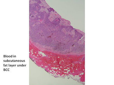
This was the pathology of the top shave
This is the histology of the second shave of the pigmented area.
Dr Ian McColl said...
I have never had another case like this one! There was not a hint of pigmentation in the top part of this BCC. It appears to have a thick infiltrating histology superiorly with a nodular pigmented BCC underneath, plus the bit of haemorrhage as you say. I thought the enlarged dermatoscopic view did show some linear serpentine vessels in the blue grey area.
5:46 PM
Dr Ian McColl said...
You might be wondering what I did with him. Well at the time after the second deep shave I just completed curreting the rest of it. I saw him 3 months later and it had all healed nicely by secondary intention. The skin was supple without evidence of infiltrating BCC. He is aged 89 and I really thought a big wedge was a bit much to carry out.Will keep an eye on it though.


No comments:
Post a Comment
Note: Only a member of this blog may post a comment.